Department of Radiodiagnosis
- Introduction of Department
- Vision
- Facilities of Department
- Faculty Members
- Faculty Residents
- Courses
- Department Publications
Introduction
Department of Radio-Diagnosis is functioning since the inception of college, in the earlier years the department used to function in the Zonal Hospital, Dharmshala, with the scarcity of the space and infrastructure, still computed tomography and sonography facilities were available. The hospital shifted to the campus build in Tanda in 2007-08 and began providing routine as well as emergency radiology services i.e. USG, color Doppler, CT, MRI, interventional image guided procedure, X-rays and fluoroscopic are being provided to the public. The department is situated in the ground floor of treatment block of the hospital. Almost all the present day imaging modalities are available in the department. We have been doing almost all image guided interventional radiological procedures in the department. We started with the DNB Radio-Diagnosis course in 2002 with two students which gradually increased to six students being admitted at present in addition we are running MD Radio-Diagnosis (recognized) admitting five student per year. We are also running B.Sc. radiology courses in the department admitting 10 students per year. We are also training the MBBS student in the radiology as per latest MCI teaching module giving them requisite exposure in USG, CT and MRI.
We have been carrying out research work in the department and publishing the research work in the journals of national and international repute. We are exposing the faculty and residents to the various CMEs throughout country and also been organizing conferences and training programmers in the institute itself. We are growing leap and bound as a department and trying our best to keep pace with the ever growing field of radiology. All this is possible due to continuous and sustained efforts of previous Heads of the Department; Dr. NK Kaushik, Dr. DS Dhiman, Dr. SL Sharma, Dr, Asha Negi and Dr. RG Sood.
Vision
Vision
Vision of the Department of Radiology, Dr RPGMC Tanda is to be one of the best Departments in the country in providing timely, cost-efficient, and high quality Medical Imaging and Image-guided therapy services for a diverse Patient population. Our Department will also play a major and vital role in the education of Patients, Trainees, Healthcare providers, Healthcare Administrators conveying the important and critical function that Medical Imaging and Image-guided therapy serves in improving the outcomes and advancing the care of Patients. In addition, our Department will continue to make significant contributions to the discovery of innovative Imaging technologies and Image-guided therapy that make a difference for the enhancement of Healthcare delivery. Our Department will learn from, build on and respect the past, be active and aggressive in improving the present, but look to the future, and rather than asking “why," we will ask “why not."
Mission
The Mission of the Department of Radiology and Medical Imaging at Dr RPGMC Kangra at Tanda is to provide compassionate, caring, and high quality Medical Imaging and Image-guided therapy services to improve the quality of life for our Patients and their families. Our leadership role in the scientific advancement of Medical Imaging and Image-Guided therapy services in a cost-efficient, less invasive and safe manner, while educating our referring Physicians, Physicians-in-training, medical students, allied health professionals remains critical to our Mission. Our Research and Scholarly activities will continue to generate innovative solutions and new knowledge that makes a difference for Patient care and the future of Healthcare.
Strategic Goals
Immediate goals is recognition of MD degree recognised by the MCI
For this equipment requirement is:
1. Biplane DSA System.
2. One Fluoro digital System.
3. Two digital mobile X-ray Machines
Patient Care
• To be recognized as offering the highest quality subspecialty Medical Imaging services and best value in our region and nationally, comparable to or better than any well-respected Medical Imaging Healthcare organization
• To add significant value to Patient care and the overall Healthcare system and to effectively manage imaging resources
• To provide Patients the best possible clinical care, using Medical Imaging technology and Image-guided therapy, in a caring, safe, high quality, cost-efficient, and timely manner
To provide organ-based, sub-specialized, and world-class consultations for Medical Imaging and Image-guided therapy services for Patients, referring Physicians, the Healthcare providers
• To provide Patients and referring Physicians access to our services via traditional on-site referrals, over-reading of films obtained at outside facilities, and tele-radiology
• To educate Patients, referring Physicians, Hospital Administrators and improve their understanding of both the hazards and benefits of Medical Imaging and Image-guided therapy
• To communicate with Patients, referring Physicians other Healthcare providers the best and most cost-effective Medical Imaging and Image-guided therapy for Patient specific questions being addressed
• To provide the data and protocols to optimize the implementation and use of new technology
• To develop and meet metrics for timely Patient access to Medical Imaging and Image-guided therapy
• To establish an infrastructure to support efforts for continuous quality improvement and patient care quality and safety initiatives by identifying and validating quantitative measures for objective outcomes assessment and comparisons to best practice models
• To lead and serve on Departmental, Institutional, Community, National, and International Medical organizations on clinical initiatives
• To be Locally, Regionally, Nationally and Internationally recognized as an outstanding clinical Department
• To create an infrastructure to support effective and efficient clinical activities
• To develop and implement focused outcomes measures for clinical activities and initiatives
• To establish a forum for frequent and effective Divisional and intra-Departmental communications around clinical activities and initiatives
Education
• To educate medical students, residents, fellows, instructors, researchers, faculty and allied health professionals in the field of Imaging and Image-guided therapeutic services by employing opportunities provided in the clinical care arena, multidisciplinary teaching and case review conferences, didactic lectures, informal sessions, and research seminars so trainees and Healthcare professionals can develop a broad foundation of knowledge upon which to base their future careers
• To lead and serve on Departmental, Institutional, Community, National, and International Medical organizations educational initiatives
• To be Locally, Regionally, Nationally and Internationally recognized as an outstanding teaching Department
• To be available and provide the necessary educational information to Hospital Administrators
• To develop and implement outcomes measures for educational efforts
• To create an infrastructure to support effective and efficient educational activities
• To establish a forum for frequent and effective Divisional and intra-Departmental communications around educational initiatives
• To develop and support efforts to promote Faculty development and mentoring
• To develop and implement educational curricula with focused and objective outcomes measures
Research
• To discover new medical knowledge and synthesize and refine existing medical knowledge to improve the clinical practice of Imaging and Image-guided therapy for the Healthcare system
• To support the advancement of the Imaging sciences and Image-guided, minimally invasive therapy and technologies through basic and clinical research in an effort to minimize Patient morbidity and costs, improve the quality of Patient care, and enhance the quality of life for our Patients
• To lead and serve on Departmental, Institutional, Community, National, and International Medical organizations on research initiatives
• To be Locally, Regionally, Nationally and Internationally recognized as an outstanding Department for Imaging research and scholarly activity
• To be available to and participate with Funding organizations and Legislators involved with providing support for research activities
• To create an infrastructure to support effective and efficient research and scholarly activities;
• To develop and implement outcomes measures for research and scholarly initiatives
• To establish a forum for frequent and effective Divisional and intra-Departmental communications around research and scholarly initiatives.
Facilities
The Department is one of the best equipped department in the institute. Following images modalities are available:
• X-Ray
• Image Intensifier
• Ultrasongraphy
• Color Doppler
• Mammography
• 128 Slice CT
• 1.5 Tesla MRI
• DSA Installed under department of Radio-Diagnosis installed in Department of Cardiology.
Faculty Members
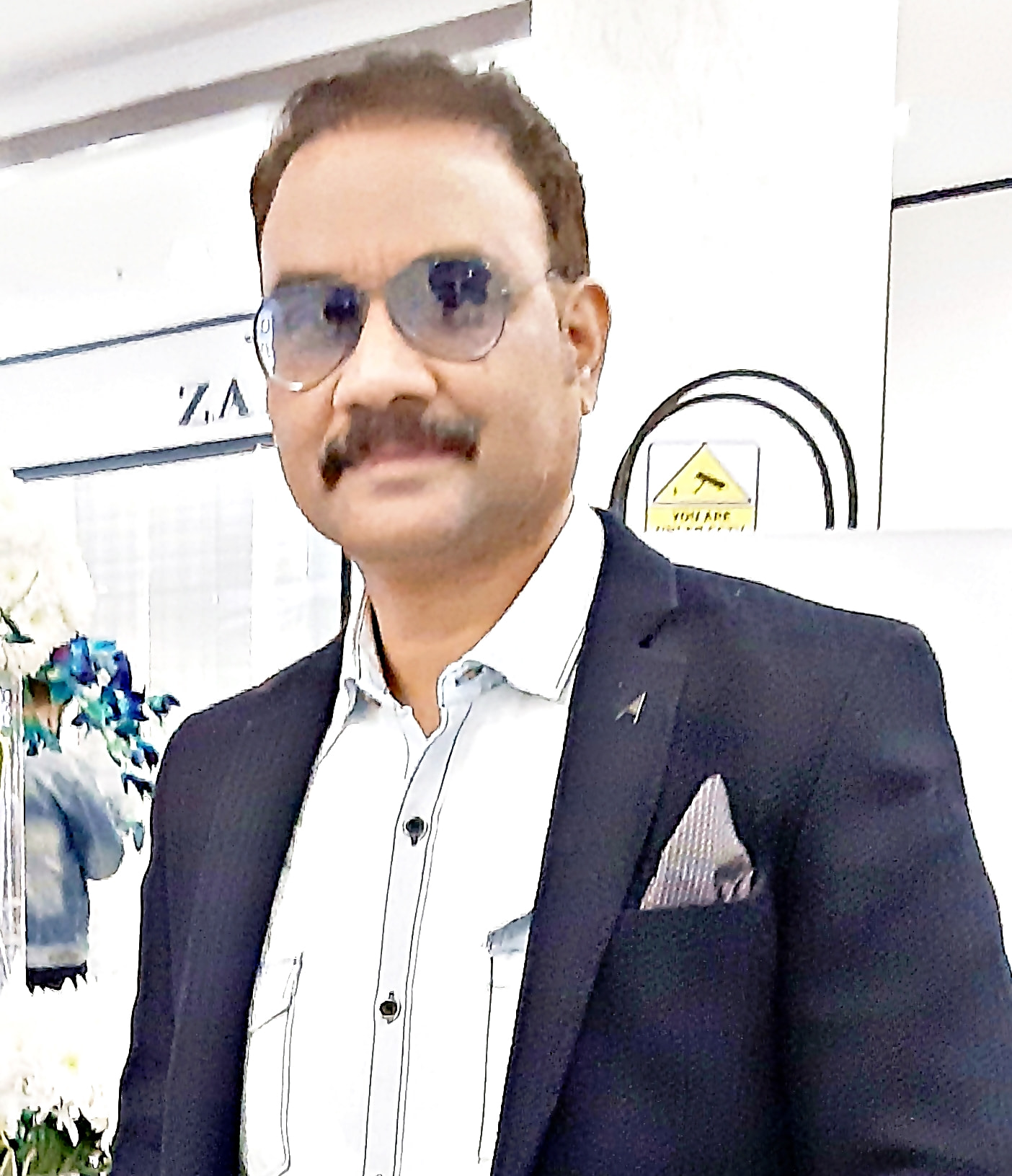
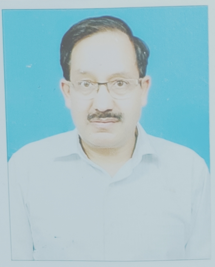
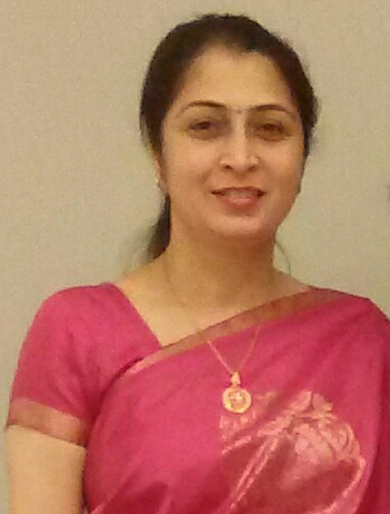
| # | Photo | Name | Qualifications | Designation | Registration Number | Email ID | Date of Joining | Awards |
|---|---|---|---|---|---|---|---|---|
| 1 |  |
Dr. Narvir Singh Chauhan | Professor and Head | drnarvir@gmail.com | 01-05- 2008 | |||
| 2 |  |
Dr.Dinesh Sood | Professor | Sud.dineshsa@gmail.com | ||||
| 3 |  |
Dr.Preeti Takkar Kapila | Assistant Professor | preetitakkar@yahoo.co.in | 06-02- 2021 | |||
| 4 |  |
Dr Nishant Nayar | Assistant Professor | nishant23rpgmc@gmail.com | 02-11- 2018 |
Senior Residents
| # | Name | Qualification | Date of Joining |
|---|---|---|---|
| 1 | Dr.Vijay | 10-09-2020 | |
| 2 | Dr.Pradeep | 27-12-2020 | |
| 3 | Dr.Ankur | 30-12-2020 | |
| 4 | Dr.Akshay | 16-03-2022 | |
| 5 | Dr.Ashish | 23-03-2022 | |
| 6 | Dr.Shikha | 22-03-2022 |
Junior Residents
| # | Name | Date of Joining |
|---|---|---|
| 1 | Dr.Ankita | 09-07-2020 |
| 2 | Dr.Pankaj | 25-07-2020 |
| 3 | Dr.Shalini | 30-07-2020 |
| 4 | Dr.Archita | 30-07-2020 |
| 5 | Dr.Arpit | 19-08-2020 |
| 6 | Dr.Pooja Kumari | 10-03-2022 |
| 7 | Dr.Gowripattapu Sirisha | 10-03-2022 |
| 8 | Dr.Ankush Kumar | 10-03-2022 |
| 9 | Dr.Rishav Dhiman | 05-11-2022 |
| 10 | Dr.Ameesh Thakur | 05-11-2022 |
| 11 | Dr.Gajulapalli Hima Bindu | 08-11-2022 |
S.O.P's
Teaching Roster
Emergency duty roster
Courses
- MBBS
- MD (Radiodiganosis)
- B.Sc. (Radio Diaganosis & Imaaging)
Labs
| # | Name | Number of labs |
|---|
Syllabus
Competency based under graduate curriculum
Department Highlights (Past 5 Years)
Publication List:
Kaur T, Sood D, Chauhan NS, Soni P. MRI evaluation of brain in paediatric patients presenting with developmental delay. Poster. European Congress of Radiology-ECR 2018 DOI:10.1594/ecr2018/C-1084 DOI-Link:https://dx.DOI.org/10.1594/ecr2018/C-1084
Chauhan NS, Chauhan IS, Sharma M, Rana L. Comparison of low dose protocol of MDCT with routine dose protocol in diagnostic evaluation of lower extremity fractures. Poster. European Congress of Radiology-ECR 2019 C-1046 DOI10.26044/ecr2019/C-1046. DOI link: https://dx.DOI.org/10.26044/ecr2019/C-1046
Chauhan NS, Kapila PT, Sharma M Forget me not': A pictorial essay on the diverse spectrum of some rare or lesser known pathologies affecting the soft tissues. Poster. European Congress of Radiology-ECR 2019 C1047. DOI: 10.26044/ecr2019/C-1047 DOI-Link:https://dx.DOI.org/10.26044/ecr2019/C-1047
Rana L, SoodD ,Chauhan NS, Gurnal P. MRI Imaging of Complicated Simple Bone Cyst. EC Orthopaedics 10.3 (2019).
Rana L, SoodD ,Chauhan NS EC Orthopaedics Case Report Diastematomylia on MRI -Case Report. February 2020. 11.4 (2020): 59-61.
Rana L, Sood D , Chauhan NS, Gurnal P, Nayyar N, Singh D. Singh Medialisation of internal carotid artery in the neck-acase report of Radiology.MOJ Proteomics Bioinform. 2020;9(2):56‒57.DOI: 10.15406/mojpb.2020.09.00279
Pandov P, Sharma A, Chauhan NS. Case Report: Imaging In A Case Of Malignant Ovarian Serous Cystadenocarcinoma With Features Of Peritoneal Carcinomatosis. December 2021 International Journal of Current Research 2019;11(11):8156-8159. https://DOI.org/10.24941/ijcr.37126.11.2019
Kumar S, Shivbrat Sharma S, Chauhan NS. Endoscopic Assisted Infra-cochlear Approach for Excision of a Large Congenital Hypo-tympanic Cholesteatoma: A Case Report. Oman Medical Journal.January 2022. DOI: 10.5001/omj.2023.37
Kumar V, Gupta N, Ambekar MM, Sharma N, Sood P, Sharma V, Chauhan NS, Sharma R, Mahajan S. Agenesis of dorsal pancreas causing extra hepatic portal vein obstruction in a patient of symptomatic cholelithiasis: a case report. International Surgery Journal. 2018 Jun 25;5(7):2682-4. DOI: 10.18203/2349-2902.isj20182799
Sharma B, Raina S, Sharma R, Chauhan NS. Double trouble: Cerebral vein thrombosis in a young female with ulcerative colitis. International Journal of Health and allied sciences 2015;4:28-29DOI: 10.4103/2278-344X.149252
Raina S, Kaul R, Kaur N, Chauhan NS. Eosinophilic granulomatosis with polyangitis (Churg–Strauss syndrome): a diagnostic rarity with an atypical presentation.Egyptian Journal of Rheumatology and Rehabilitation. 2014;41:187- 189cDOI: 10.4103/1110-161X.147363
Rana L, Sood D, Diwedi A, Parminder S, Sahu SK , Chauhan NS. Portal Annular Pancreas with Portal Cavernoma Formation with Associated Dorsal Pancreatic Agenesis - A Rare Case Report 2018; 1(2):10-13 https://DOI.org/10.31579/2642- 1674/007
Rana L , Sood D, Verma N, Parminder S, Sahu SK and Chauhan NS. Cryptococcoma’s on Mr Imaging-A Case Report. Medical Research and Clinical Case Reports 2018;2(3)
Rana L, Sood D, Chauhan NS, Gurnal P, Manjuswamy HR Computed Tomography Imaging of Sealed Perforated Diverticulitis-A Rarely Encountered Entity. Acta Scientific Medical Sciences. 2019;3:68-9.
Nayyar N, Sood D, Chauhan NS, Kumar N, Thakur M, Thakur D. Spontaneous subcapsular renal haematoma in a diabetic patient with acute pyelonephritis. Tropical Doctor. 2021 May 27:00494755211017019. DOI: 10.1177/00494755211017019
Sharma A, Chauhan NS, Pandoh P Sharma D. Frontonasal Epidermoid Cyst With Patent Dermal Sinus Tract Opening On The Dorsum Of Nose And Intracranial Extension Through Cribriform Plate Defect: A Rare Case Report With Review Of Literature Int. J. of Adv Research (IJAR). Feb 2021;9: 687-690
Rana L, Sood D, Chauhan NS,Gurnal P, Sharma KS, Negi P, SharmaA..Celiac Trunk Abnormalities Presenting with Intractable Epigastric Pain-Case Reports.Journal of Medical Research and Case Reports 2020;2(2)
Lokesh L, Dinesh S, Chauhan NS, Pooja G and Manjuswamy HR. MRI Imaging of Spinal Intramedullary Tuberculoma.ClinRadiol Imaging J. December 2018;2(4):000135DOI: 10.23880/CRIJ-16000135
Rana L, Sood D, Chauhan NS,Gurnal P and ManjuswamyHR.CysticEncephalomlacia-Mr Imaging. Biomed J Sci& Tech Res. Jan 2019; 12(4):1-2DOI: 10.26717/BJSTR.2019.12.002277
Rana L, Sood D, Chauhan NS,Gurnal P and Manjuswamy HR. MRI Imaging of Osmotic Demyelination Syndrome. Biomed J Sci& Tech Res. Dec 2018DOI: 10.26717/BJSTR.2018.12.002246
Rana L, Sood D, Chauhan N, Gurnal P, Manjuswamy HR. MRI Imaging of Craniopharyngioma. Ann Clin Case Rep. 2019; 4.;1576.
Dev M, Sharma M, Shukla R, Sood D, Chauhan NS. Isolated Agenesis of Right Upper Lobe of Lung: A Case Report. American Journal of Diagnostic Imaging. 2021;7(1):1-4. DOI: 10.5455/ajdi.20191119051748
Sharma M, Sood D, Chauhan NS, Dev M. US of Right Upper Quadrant Pain: Some Uncommon but Noteworthy Causes. RadioGraphics. 2018 Nov;38(7):2212-3. DOI: 10.1148/rg.2018180190
Goyal VD, Kumar S, Chauhan N, Shukla A, Kaul R. Mesenteric Lymph Node Hamartoma (Castleman's Disease) in Association with Superior Mesenteric Arteriovenous Fistula. J ClinDiagn Res. 2014 Dec;8(12):ND05-6. DOI 10.7860/JCDR/2014/10849.5327.
Chander B, Dogra SS, Kaul R, Preet K, Sharma R, Chauhan NS. Lymphangiomatosis: Two cases with unique presentations, salience of nomenclature, and diagnosis. J Cancer Res Ther. 2015 Jul-Sep;11(3):652. DOI: 10.4103/0973-1482.139401
Nimkar K, Sood D, Soni P, Chauhan N, Surya M. Giant Atretic Occipital Lipoencephalocele in an Adult with Bony Outgrowth. Pol J Radiol. 2016 Aug 21;81:392-4. DOI: 10.12659/PJR.895453. PMID: 27617049; PMCID: PMC4994857.
Sharma M, Sood, D; Chauhan N; Negi P. Acute necrotizing encephalopathy of childhood. Neurology India; Mumbai Vol. 67, Iss. 2, (Mar/Apr 2019): 610-611. DOI:10.4103/0028-3886.257990
Shekhar S, Chauhan NS, Singh K, Surya M. Delayed and successful manual removal of abnormally adherent placenta (AAP) necessitated by uterine sepsis following conservative management with adjuvant methotrexate: A rewarding clinical experience. South African journal of obstetrics and gynaecology 2103 19(1):19-21 DOI: 10.7196/SAJOG.570
Shandil A, Verma N, Sood D, Chauhan NS, Bhardwaj A, Chauhan R, Shandil A. Comparison of the findings of gray scale sonography, colour Doppler and CT angiography in evaluation of Extracranial Carotid vascular stenosis.SJMSCR. June 2019. 541-549 : DOI: https://dx.doi.org/10.18535/jmscr/v7i6.92
Sharma M, Sood D, Chauhan NS, Manjuswamy RH, Kapila PT. Pictorial Essay: Classic Signs in Pediatric Neuroradiology. Current pediatric reviews. 2019 Sep 16;16(1):6-16. DOI: 10.2174/1573396315666190916141023
Sharma M, Sood D, Chauhan NS, Verma N, Kapila P. Inferior right hepatic vein on routine contrast-enhanced CT of the abdomen: prevalence and correlation with right hepatic vein size. Clinical radiology. 2019 Sep 1;74(9):735-e9. DOI: 10.1016/j.crad.2019.05.023
Raina S, Mahesh D, Rajendra G, Chauhan NS. Reversible posterior leukoencephalopathy syndrome. J Neurosci Rural Pract. 2012 May;3(2):222-4. DOI: 10.4103/0976-3147.98262. PubMed PMID: 22865991
Bhoil R, Ahluwalia A, Chauhan NS. Herlyn Werner Wunderlich Syndrome with Hematocolpos: An Unusual Case Report of Full Diagnostic Approach and Treatment. Int J FertilSteril. 2016 Apr-Jun;10(1):136-40. Epub 2016 Apr 5. DOI: 10.22074/ijfs.2016.4779
Sood D, Soni PK, Chauhan NS, Rana L, Nautiyal H, Kanue YS. Role of Magnetic Resonance Imaging in Diagnosing Patients Presenting with Non-Traumatic Hip Pain at a Tertiary Care Hospital in Sub-Himalayan Region. Annals of International Medical and Dental Research.;4(4):10. DOI: 10.21276/aimdr.2018.4.4.RD3
Sood D, Chauhan NS, Sharma G. Role of B scan ultrasonography in evaluating orbital pathology June 2018IOSR Journal of Dental and Medical Sciences 17(1)DOI: 10.9790/0853-1701173235
Sood D, Chauhan N S, Kumar H. Evaluation of Scrotal Diseases with High Frequency Ultrasonography and Colour Doppler Sonography. June 2018 IOSR Journal of Dental and Medical Sciences 16(12)DOI: 10.9790/0853-1612092831
Kaur T, Chauhan NS.MRI evaluation of brain in paediatric patients presenting with developmental delay.2018. International Journal of Current Advanced Research 2018 June ;7(6):13137-40 DOI: 10.24327/ijcar.2018.13140.2329
Awasthi B, Raina SK, Negi V, Chauhan NS, Kalia S. Morphometric study of lower end femur by using helical computed tomography. Indian journal of orthopaedics. 2019 Apr;53(2):304-8.DOI: 10.4103/ortho.IJOrtho_136_17
Awasthi B, Raina SK, Chauhan N, Sehgal M, Sharma V, Thakur L. Anthropometric characterisation of elbow angles and lines among Indian children. Advances in Human Biology. 2017 May 1;7(2):71.DOI: 10.4103/AIHB.AIHB_2_17
Sehgal M, Awasthi B, Raina SK, Chauhan N, Sharma V, Thakur L. Anthropometric characterisation of elbow angles and lines among Tibetan children (3–13 years) seeking refuge in India: A comparison survey. Advances in Human Biology. 2018 Jan 1;8(1):28. DOI: 10.4103/AIHB.AIHB_18_17
Awasthi B, Raina SK, Chauhan NS, Sehgal M, Sharma V, Thakur L. Distribution of elbow injuries among children 3-13 years of age in North-West India. Tropical journal of Medical research 2016;19:61-63 DOI: 10.4103/AIHB.AIHB_18_17
Kumar S, Joshi A, Tuli R, Chauhan NS.Traumatic Optic Neuropathy Our Experience with Combined Therapy. Indian Journal of Neurotrauma December 2021. (online) DOI: 10.1055/s-0041-1739479
Kalia S, Chauhan NS, Sood D, Singh I. MDCT evaluation of fractures injuries of joints in patients with lower extremity trauma: A single institution prospective observational study. International Journal of Orthopaedics. 2020;6(1):663-7. DOI: 10.22271/ortho.2020.v6.i1l.1940
Chauhan NS, Kumar S, Chander B, Sharmal S, Garg S; Xanthogranulomatous cholecystitis: A rare radiological diagnosis. Sub Himalayan Journal of Health Research.
Chauhan NS, Kaul R, Thakur S, Nimkar K. Unusual case of sciatic nerve and deep pelvic endometriosis with lumbosacral plexus spread presenting with muscular atrophy and foot drop. Archives of Medicine and Health Sciences. 2017 Jan 1;5(1):79. DOI: 10.4103/amhs.amhs_128_16
Chauhan NS. A 48-year-old Man with Epigastric Pain and Melena. Emerg (Tehran). (Continues as archives of Academic Emergency Medicine) 2016 Nov;4(4):211-213. PMID: 27800543; PMCID: PMC5007914. DOI: 10.22037/aaem.v4i4.253
Chauhan NS. JAMA Intern Med. 2016;176(7):889. DOI:10.1001/jamainternmed.2016.2787. Article July 2016JAMA Internal Medicine
Chauhan NS, Verma A, Sharma S, Garg S. A rare case of non-puerperal uterine inversion with herniation of both fallopian tubes in the inversion cleft. Medical Journal Armed Forces India. 2020 Jul;76(3):349. DOI: 10.1016/j.mjafi.2018.11.001
Chauhan NS, Singh S. A queer case of Parry–Romberg syndrome with coexisting psychiatric ailment. Indian journal of psychiatry. 2019 Jan;61(1):97. DOI: 10.4103/psychiatry.IndianJPsychiatry_340_17
Chauhan NS, Sharma Y; Sonographic diagnosis of infected chronic uterine inversion at 12 weeks postpartum Ultrasound (British Medical Ultrasound Society) 2011;19(4):227-2292.https://DOI.org/10.1258/ult.2011.011014
Chauhan NS, Bharti R, Chander B, Kumar S. Pediatric Clear Cell Renal Cell Carcinoma with Pelvicalyceal System Invasion and Contiguous Extension Upto Bladder: Novel and Bizarre Tumor Behaviour. Pol J Radiol. 2016 May 29;81:256- 60. DOI: 10.12659/PJR.896088
Chauhan NS, Sharma YP. Dual thyroid ectopia: A case report. Journal of medical imaging and radiation oncology 2011;55(4); 404-406. DOI: 10.1111/j.1754- 9485.2011.02289.x
Chauhan NS, Banday IA, Morey P, Deshmukh A. External brain tamponade: a rare complication of decompressive craniectomy. Intern Emerg Med. 2016 Mar 16. [Epub ahead of print] PubMed PMID: 26983955. DOI: 10.1007/s11739-016-1436-4
Chauhan NS, Sood D. Case report: Colonic bezoar due to Box Myrtle seeds: A very rare occurrence. Indian J Radiol Imaging. 2011 Jan;21(1):21-3. DOI: 10.4103/0971-3026.76049. PMID: 21431028
Chauhan NS, Sharma M, Bhardwaj A. Classical case of late-infantile form of metachromatic leukodystrophy. J Neurosci Rural Pract. 2016 Jul-Sep;7(3):473-5. DOI: 10.4103/0976-3147.181482
Chauhan NS, Sharma YP. A child with skin nodules and extensive soft tissue calcification. British Journal of Radiology. 2012 Feb;85(1010):193-5. DOI: 10.1259/bjr/95392214
Chauhan NS; Sharma, YP; Sharma S; Sharma S.Complete androgen insensitivity syndrome with microtia: a rare presentation. Japanese journal of radiology.2011;29(1)67-71DOI: 10.1007/s11604-010-0501-6
Chauhan NS, Gupta A, Soni PK, Surya M, Mahajan SR. Iatrogenic uterine perforation with abdominal extrusion of fetal parts: a rare radiological diagnosis. J Radiol Case Rep. 2013 Jan;7(1):41-7. DOI: 10.3941/jrcr.v7i1.1332. Epub 2013 Jan 1
Chauhan NS, Kumar S, Bhoil R. Rapunzel Syndrome: Rare 'Tale' of a Broken'Tail'. Pol J Radiol. 2016 Apr 14;81:166-9. DOI: 10.12659/PJR.896154
Chauhan NS, Sharma YP, Bhagra T, Sud B. Branchial fistula arising from pyriform fossa: CT diagnosis of a case and discussion of radiological features. Clin Imaging. 2012 Sep-Oct;36(5):591-4. DOI: 10.1016/j.clinimag.2012.01.032. Epub 2012 Jun 8.
Chauhan NS, Kumar S. Torsion of a Wandering Spleen Presenting as Acute Abdomen. Pol J Radiol. 2016 Mar 15;81:110-3. DOI: 10.12659/PJR.895972. PMID: 27057261; PMCID: PMC4795092.
Chauhan NS, Sharma YP, Bhagra T, Sud B. Persistence of multiple emissary veins of posterior fossa with unusual origin of left petrosquamosal sinus from mastoid emissary. Surgical and radiologic anatomy 2011;33(9):827-831DOI: 10.1007/s00276-011-0822-x
Chauhan NS, SoniPK,ManjuswamyHR. A Systematic Analysis of Medicolegal Radiological Investigations and Quantification of the Burden of Court Evidences on Radiology Department of a Tertiary Level Teaching Institute of North India. Indian Journal of Forensic Medicine & Toxicology. 2019 Jan 1;13(1).DOI: 10.5958/0973- 9130.2019.00015.X
Mahajan VK, Chauhan NS, Rana BS, Mehta KS, Hooda S, Chauhan PS, Kukreja A. The association between chronic plaque psoriasis and non-alcoholic fatty liver disease in Indian patients: Results of a pilot study. Journal of Clinical and Experimental Hepatology. 2021 Dec 3. DOI: 10.1016/j.jceh.2021.11.016
Chauhan NS, Thakur L, Singh S, Awasthi B, Raina SK. Morphometric evaluation of acetabular dimensions: A computed tomography-based study from a tertiary care center in rural North-West India. Journal of Medical Society. 2018 Sep 1;32(3):195.DOI: 10.4103/jms.jms_48_17
Chauhan NS, Sood D, Shukla A. Magnetic Resonance Imaging (MRI) Characterization of Perianal Fistulous Disease in a Rural Based Tertiary Hospital of North India. Pol J Radiol. 2016 Dec 22;81:611-617. DOI: 10.12659/PJR.899315. PMID: 28096904; PMCID: PMC5201120.
Chauhan NS, Ahluwalia A, Sharma YP, Thakur L. A prospective comparative study of high resolution ultrasound and MRI in the diagnosis of rotator cuff tears in a tertiary hospital of North India. Polish journal of radiology. 2016;81:491.DOI: 10.12659/PJR.897830
Chauhan NS, Sood D, Takkar P, Dhadwal DS, Kapila R. Quantitative assessment of airway and parenchymal components of chronic obstructive pulmonary disease using thin-section helical computed tomography. Polish journal of radiology. 2019;84:e54.DOI: 10.5114/pjr.2019.82737
Chauhan NS; Nandolia K. Comparison of Ultrasound and MRI imaging findings in evaluation of foetal congenital anomalies: A single institution prospective observational study. Medical Journal of Armed forces of India. ISSN 0377-1237 Published online 21 January 2021. https://DOI.org/10.1016/j.mjafi.2021.12.002
Chauhan NS, Kumar S, Takkar P, Sood. Radiological Features of Rhino-Orbital- Cerebral Mucormycosis Complicating COVID-19 Illness: a Pictorial Essay. Hong Kong J Radiol. 2022;25:226-34 https://DOI.org/10.12809/hkjr2217501